"This study is helpful because it indicates that for most patients with thyroglobulin values of <0.1 ng/ml while on thyroid hormone suppression, stimulated thyroglobulin testing is unnecessary since the likelihood of identifying residual cancer is very small. These findings will cut down on the need to perform stimulated thyroglobulin testing, which is both inconvenient for patients and expensive. Long term follow up of patients with thyroid cancer, however, still requires periodic measurement of thyroglobulin, since other studies indicate that ~4% of patients with initially undetectable basal thyroglobulin levels eventually had recurrent cancer."
http://www.thyroid.org/patient-thyroid-information/ct-for-patients/vol-7-is sue-2/vol-7-issue-2-p-7-8/
When thyroglobulin is undetectable, is any further testing needed in following patients with thyroid cancer?
BACKGROUND
Thyroglobulin is a protein produced by both normal and cancerous thyroid cells. Treatment of thyroid cancer frequently involves total thyroidectomy and radioiodine therapy followed by thyroid hormone therapy to suppress serum TSH and turn off any residual normal thyroid cells. In this situation, the serum thyroglobulin level can be used as a thyroid cancer marker. Indeed, if any thyroid cancer cells are present, levels of thyroglobulin are often detectable, either on TSH suppression therapy or after stimulation with rhTSH (stimulated thyroglobulin testing). Measurement of thyroglobulin under these conditions has become standard practice in the follow up of patients with thyroid cancer. This study is an analysis of many other studies as to the usefulness of measuring serum thyroglobulin levels in managing patients with thyroid cancer.
Thyroglobulin is a protein produced by both normal and cancerous thyroid cells. Treatment of thyroid cancer frequently involves total thyroidectomy and radioiodine therapy followed by thyroid hormone therapy to suppress serum TSH and turn off any residual normal thyroid cells. In this situation, the serum thyroglobulin level can be used as a thyroid cancer marker. Indeed, if any thyroid cancer cells are present, levels of thyroglobulin are often detectable, either on TSH suppression therapy or after stimulation with rhTSH (stimulated thyroglobulin testing). Measurement of thyroglobulin under these conditions has become standard practice in the follow up of patients with thyroid cancer. This study is an analysis of many other studies as to the usefulness of measuring serum thyroglobulin levels in managing patients with thyroid cancer.
THE FULL ARTICLE TITLE: Giovanella L et al, Unstimulated high-sensitive thyroglobulin in follow-up of differentiated thyroid cancer patients: a meta-analysis. J Clin Endocrinol Metab. 2013 Nov 27.
SUMMARY OF THE STUDY
This study is an analysis of many other studies evaluating the utility of measuring thyroglobulin levels under thyroid hormone suppression therapy and after stimulation with rhTSH in patients with thyroid cancer. The authors identified 9 studies that used the newer, more sensitive thyroglobulin assay. These studies included a total of 3178 patients. The investigators found that when the basal thyroglobulin level under thyroid hormone suppression therapy is <0.1 ng/ml, it accurately predicts that the stimulated thyroglobulin level will be <1, which indicates absence of residual cancer cells.
This study is an analysis of many other studies evaluating the utility of measuring thyroglobulin levels under thyroid hormone suppression therapy and after stimulation with rhTSH in patients with thyroid cancer. The authors identified 9 studies that used the newer, more sensitive thyroglobulin assay. These studies included a total of 3178 patients. The investigators found that when the basal thyroglobulin level under thyroid hormone suppression therapy is <0.1 ng/ml, it accurately predicts that the stimulated thyroglobulin level will be <1, which indicates absence of residual cancer cells.
WHAT ARE THE IMPLICATIONS OF THIS STUDY? This study is helpful because it indicates that for most patients with thyroglobulin values of <0.1 ng/ml while on thyroid hormone suppression, stimulated thyroglobulin testing is unnecessary since the likelihood of identifying residual cancer is very small. These findings will cut down on the need to perform stimulated thyroglobulin testing, which is both inconvenient for patients and expensive. Long term follow up of patients with thyroid cancer, however, still requires periodic measurement of thyroglobulin, since other studies indicate that ~4% of patients with initially undetectable basal thyroglobulin levels eventually had recurrent cancer.
—M. Regina Castro, MD
ATA THYROID BROCHURE LINKS
Thyroid cancer: http://www.thyroid.org/cancer-of-the-thyroid-gland
Radioactive Iodine Therapy: http://www.thyroid.org/radioactive-iodine

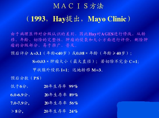 点击放大图片
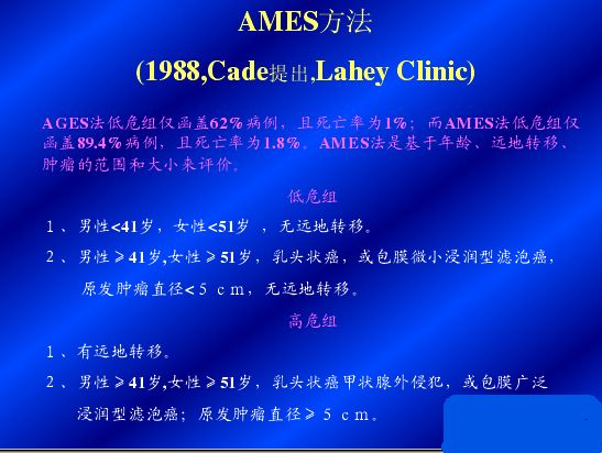 点击放大图片
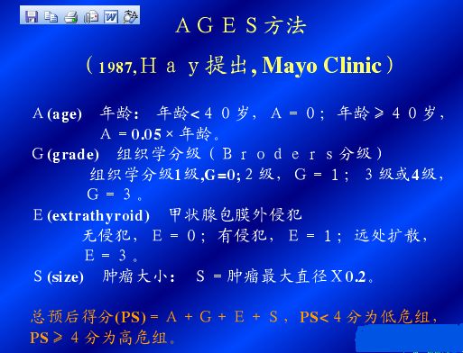 点击放大图片